- Visibility 215 Views
- Downloads 27 Downloads
- Permissions
- DOI 10.18231/j.ijca.2023.018
-
CrossMark

Anesthetic management of a Kyphoscoliotic parturient with Mitral valve prolapse posted for emergency cesarean section
Abstract
Kyphoscoliosis is a spinal deformity with forward and lateral deviation of the normal spine. It occurs in up to 4% of the population. This condition in pregnancy causes altered growth of gravid uterus and leads to unpredictable delivery. These deformities if extreme can cause obstructed labor which will require cesarean section and are usually associated with physiologic derangements in cardiopulmonary function causing difficulties in conducting both general and regional anaesthesia. One of the causes of kyphoscoliosis can be secondary to cardiothoracic surgery in the childhood. Subarachnoid block in kyphoscoliotic patients is always challenging to an anesthesiologist due to the deformity of spine and spread of anesthetic drug to unpredicted level. We present a known case of post cardiac surgery, mitral valve prolapse with kyphoscoliosis in the thoracolumbar region posted for emergency cesarean section managed under subarachnoid blockade.
Introduction
Kyphoscoliosisis defined as a deviation of the curvature of the spine, both in the sagittal and coronal planes, also including rotation of the spinal axis[1] with Cobb angle of more than 40 degrees in the thoracolumbar spine.[2] The incidence of idiopathic kyphoscoliosis is around 4 per 1000 population with female to male ratio of 4:1.[3] Etiologies being secondary to causes including degenerative and senile changes, diseases associated with inflammation, post-traumatic fractures, iatrogenic post-surgical changes and repeated microtrauma.[4] Other mechanisms also contribute like congenital vertebral anomalies, thoracogenic causes and the presence of syndromic conditions. Sternotomy and thoracotomy are also associated with the development of scoliosis.[5], [6], [7], [8]
Problems with kyphoscoliosis are difficult airway, restrictive pulmonary disease and decreased cardiac output.[9], [10] Central neuraxial blocks have anesthetic challenge due to difficulties in palpating anatomical landmarks, performing dural puncture and difficulty in predicting the extent of block.[11], [12], [13] Several case reports show that spinal anesthesia is safe in these set of patients.[14] We may encounter such cases of kyphoscoliosis posted for cesarean section in our day to day practice.
Case Report
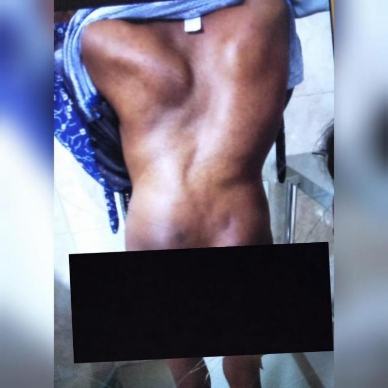
An unbooked 23year old kyphoscoliotic primigravida with 38 weeks of gestation with height and weight of 123cm and 32kg respectively with history of corrective cardiac valve surgery in her childhood was posted for emergency cesarean section. The cardiac surgery was done 17 years back with no previous records available. She had no other known comorbidities and gave no history of breathlessness throughout her antenatal period. Airway assessment showed anadequate mouth opening with Mallampatti of Grade 2, normal dentition, normal temper mandibular joint and neck movements . On examination of the spine there was thoracic kyphoscoliosis with winging of scapula. Her blood investigations were within normal limits with hemoglobin of 11.2 gm% and a normal ECG. She had taken her last meal 8 hours ago.
Her baseline vitals were examined and were as follows: pulse rate of 83 beats/min which was regular, blood pressure of 110/80mm Hg in left lateral position and Spo2 of 98% on room air. On respiratory examination, normal bilateral vesicular breath sounds were heard allover the chest. On cardiovascular examination, systolic murmur was heard over mitral area with rest of the systemic examination normal. Her 2D ECHO showed post cardiac surgery status and MVP with mild MR, no ASD/ VSD/ PDA ,PASP -30mmHg, EF- 60%. We planned to conduct cesarean section under spinal anaesthesia as it was an unbooked case with awaited RTPCR report and regional anesthesia weighed more beneficial than general anesthesia in this case in terms of anticipated difficult airway and anesthetist’s safety during the COVID pandemic. In the preoperative room, patient was secured with an 18G IV cannula over dorsum of left hand and preloaded with 5 ml/kg of Ringer lactate. Injection Ranitidine 50mg IV and Injection Metoclopramide 10mg IV were given as premedication as apart of aspiration prophylaxis. Machine was checked and difficult airway cart was kept ready. Patient was shifted to the operation table and all standard multi channel parameters like continuous electrocardiogram, non invasive blood pressure and pulse oximetry were connected and baseline readings were taken. With patient in left lateral position, under aseptic precautions lumbar puncture was done using the 25 G Quincke Babcock spinal needle at level L3 -4 space through paramedian approach as midline approach was difficult. After confirming back flow of CSF, local anesthetic Injection Bupivacaine heavy 0.5% 1mlplus Fentany l12.5 micrograms was given intrathecally and patient was made to lie supine and supplemental oxygen 5 litres per minute was administered through face mask. Once the sensory level of T6 was achieved, surgeon was asked to start the surgery. Vitals were recorded in the intraoperative period at regular intervals and the patient was stable throughout the surgery. She was then shifted to post-operative ward for further management.
Discussion
Scoliosis is obtained from the greek word meaning ‘crooked’. Patients with scoliosis have restrictive lung disease which decreases vital capacity, functional residual capacity, tidal volume and causes faster respiratory cycle1. It causes chronic hypoxia, hypercapnia, and pulmonary vasoconstriction resulting in irreversible vascular changes, pulmonary hypertension and eventually right ventricular hypertrophy and cor pulmonale. The severity of pulmonary impairment is determined by the degree of Cobb angle which is also utilized to assess severity of kyphoscoliosis, risk of progression and method of intervention.[15] The pulmonary impairment severity also depends on the number of vertebrae involved and the cephalad location of the curvature. Thoracolumbar kyphoscoliosis is very common. Moderate kyphoscoliosisis Cobb angle ranging from 25 to 100 degrees, whereas severe kyphoscoliosisis Cobb angle greater than 100 degrees.[16]
Structural abnormalities such as reduced lung volume secondary to a restrictive thoracic chest wall, decreased compliance and limited diaphragmatic movements may result in restrictive lung disease further leading to chronic hypercapnic respiratory failure and alveolar hypoventilation.[17]
Surgical correction can reduce the cardiorespiratory problems resulting in slight improvement. In our case the patient gave no history of spine surgery.
Even if the lungs are functionally normal, the distortion of the thoracic cage makes the respiratory systemless compliant and increases the work of breathing. In severe cases, displacement with rotation of the trachea and main stem bronchi may also be noted, which could cause problems during intubation for general anesthesia.[18]
Congenital heart disease (CHD) affects 1% of live births and has its association with scoliosis.[19] Current evidence revelas 10 times increase in the prevalence of scoliosis in children with CHD when compared to age-matched populations.[20] Since the patient gives history of corrective cardiac valve surgery in her childhood, this could have led to herkyphoscoliosis condition.
In our case x-ray could not be done to avoid radiological exposure to the fetus. As the case was an emergency, severity grading of the deformity and pulmonary function tests could not be done. These changes in pregnancy can worsen the respiratory function in a scoliosis patient with restrictive lung disorder. The maternal mortality and morbidity correlates well with the degree of functional impairment be for pregnancy.[1], [15] In the current scenario of covid pandemic as her RTPCR report was not available we chose to conduct cesarean section under spinal anesthesia with low dose of local anesthetic
David G Veliath et al[21] presented a case of emergency cesarean section of a 22-year-oldwoman with 39 weeks of gestation for Cephalo-pelvic disproportion with history of spine surgery at 9 years of age for correction of scoliosis for which Luque rods were placed in T4–T12 vertebrae. After discussing various modes of anesthetic options, he decided to administerspinal anesthesia due to the severe restrictive lung disease as well as full stomach of the patient. The patient was placed in lateral position and spinal anesthesia was administered at L4-5 interspace with a 26G Quinke Babcock needle using 1.8 ml of 0.5% Bupivacaine. She was monitored for 12 hours in the postoperative ward and discharged after 4 days.
Sara Korula et al[22] presented a case of childhood poliomyelitis of a 33-year-old primigravida with severe kyphoscoliosis, 32 weeks of gestation with history of grade 1 breathlessness. She was posted for emergency cesarean section due to her poor respiratory status. Patient had history of fever, cough and breathlessness for four days. To avoid postoperative hypoventilation and prolonged mechanical ventilation, case was done under epidural anesthesia. After the test dose of two percent Lignocaine and was given as graded doses over
10 min. After achieving T6 level of sensory blockade, the surgery was started with no hypotension, bradycardia in the intraoperative period. Noninvasive BIPAP was instituted in the post-operative period in ICU and was discharged after two weeks.
Dr. Chavi Seth et al[23] presented a case of 24 year old primigravida with severe thoracolumbar kyphoscoliosis with grade 1 breathlessness posted for emergency caesarean section due to non progression of labor. Past history had no known etiology for kyphoscoliosis. She developed dyspnea on exertion during second trimester, which progressively increased. Due to history suggestive of malignant hyperthermia in her siblings and known association of kyphoscoliosis with muscular dystrophies in the family, regional anaesthesia was chosen. After aseptic precautions, spinal anesthesia was administered with 25G Quincke Babcock spinal needle inL3- L4 space via paramedian approach using 1.5 ml of Bupivacaine 0.5% heavy with Injection fentany l25ug. Patient was discharged on 5th postoperative day uneventfully.
Conclusion
Spinal anesthesia is an effective and safe option for patients with kyphoscoliosis. Based on anatomical consideration in patients with scoliosis, paramedian approach with needle insertion towards convexity may offer successful lumbar puncture. Hence, patients coming for cesarean section with thoracolumbar kyphoscoliosis with mild heart disease can be safely managed under regional anesthesia after weighing the benefits and risks.
Source of Funding
Nil.
Conflicts of Interest
There are no conflicts of interest.
References
- Papaliodis D, Bonanni P, Roberts T, Hesham K, Richardson N, Cheney R. Computer Assisted Cobb Angle Measurements: A novel algorithm. Int J Spine Surg. 2017;11(3). [Google Scholar]
- Sheehan D, Grayhack J. Pediatric Scoliosis and Kyphosis: An Overview of Diagnosis, Management, and Surgical Treatment. Pediatr Ann. 2017;46(12):e472-80. [Google Scholar]
- Schwartz J, Schwartz J. Skin and musculoskeletal diseases. Anaesthesia and Coexisting. 2010. [Google Scholar]
- Ailon T, Shaffrey C, Lenke L, Harrop J, Smith J. Progressive Spinal Kyphosis in the Aging Population. Neurosurgery. 2015;77(Suppl 4):S164-72. [Google Scholar]
- Herrera-Soto J, Have K, Barry-Lane P, Myers J. Retrospective study on the development of spinal deformities following sternotomy for congenital heart disease. Spine (Phila Pa 1976). 2007;32(18):1998-2004. [Google Scholar]
- Ruiz-Iban M, Burgos J, Aguado H, Diaz-Heredia J, Roger I. Scoliosis after median sternotomy in children with congenital heart disease. Spine (Phila Pa 1976). 2005;30(8):214-8. [Google Scholar]
- Bal S, Elshershari H, Celiker R, Celiker A. Thoracic sequels after thoracotomies in children with congenital cardiac disease. Cardiol Young. 2003;13(3):264-7. [Google Scholar]
- Herrera-Soto J, Have K, Barry-Lane P, Woo A. Spinal deformity after combined thoracotomy and sternotomy for congenital heart disease. J Pediatr Orthop. 2006;26(2):211-5. [Google Scholar]
- Gambrall M. Anesthetic implications for surgical correction of scoliosis. AANA J. 2007;75(4):277-85. [Google Scholar]
- Koumbourlis A. Scoliosis and the respiratory system. Paediatr Respir Rev. 2006;7(2):152-60. [Google Scholar]
- Hebl J, Horlocker T, Schroeder D. Neuraxial anesthesia and analgesia in patients with preexisting central nervous system disorders. Anesth Analg. 2006;103(1):223-8. [Google Scholar]
- Lambert D, Giannouli E, Schmidt B. Post poliosyndrome and anesthesia. Anesthesiology. 2005;103(3):638-44. [Google Scholar]
- Chin K, Chan V, Ramlogan R, Perlas A. Real-time ultrasound-guided spinal anesthesia in patients with a challenging spinal anatomy: Two case reports. Acta Anaesthesiol Scand. 2010;54(2):252-5. [Google Scholar]
- Higashizawa T, Sugiura J, Takasugi Y. Spinal anesthesia in a patient with hemiparesis after poliomyelitis. Masui. 2003;52(12):1335-7. [Google Scholar]
- Mcmaster M, Glasby M, Singh H, Cunningham S. Lung function in congenital kyphosis and kyphoscoliosis. J Spinal Disord Tech. 2007;20(3):203-8. [Google Scholar]
- Johari J, Sharifudin M, Rahman A, Omar A, Abdullah A, Nor S. Relationship between pulmonary function and degree of spinal deformity, location of apical vertebrae and age among adolescent idiopathic scoliosis patients. Singapore Med J. 2016;57(1):33-8. [Google Scholar]
- Huang S, Wu C, Lin C, Hung C, Kuo L, Weng Y. Effect of long term intermittent nocturnal non-invasive positive pressure ventilation on patient with severe kyphoscoliosis and hypoxaemia. BMJ Case Rep. 2009;2009. [Google Scholar]
- Kulkarni A, Ambareesha M. Scoliosis and anesthetic considerations. Indian J Anaesth. 2007;51:486-95. [Google Scholar]
- Coran D, Rodgers W, Keane J, Hall J, Emans J. Spinal fusion in patients with congenital heart disease. Predictors of outcome. Clin Orthop Relat Res. 1999;364:99-107. [Google Scholar]
- Webb G, Harrison D, Connelly M. Challenges posed by the adult patient with congenital heart disease. Adv Intern Med. 1996;41:437-95. [Google Scholar]
- Veliath D, Sharma R, Ranjan R, Kumar C, Ramachandran T. Parturient with kyphoscoliosis (operated) for cesarean section. J Anaesthesiol Clin Pharmacol. 2012;28(1):124-6. [Google Scholar]
- Korula S, Ipe S, Abraham SP. Parturient with severe kyphoscoliosis: Ananesthetic challenge. J Obstet Anaesth Crit. 2011;1:81-4. [Google Scholar]
- Sethi C, Kumar R, Srivastava S. Anaesthetic management of kyphoscoliotic gravida scheduled for emergency caesarean section. Int J Adv Res Biol Sci. 2016;3(8):132-5. [Google Scholar]