Case History
36 years old, female patient, case of achondoplastic dwarfism had a uterine fibroid with heavy per vaginal bleeding posted for total abdominal hysterectomy. She stands 100cm and weighted 33 kg (BMI-33kg/m2). Patient had dyspnoea on exersion MMRC grade I since childhood. Achondroplasia was associated with thoracolumbar kyphoscoliosis, fused cervical spine, short neck and restricted neck movements with mild pulmonary restrictive disease. She did not have any significant past history of chronic illness or any surgeries.
On general examination, she was afebrile with pulse of 90/min, blood pressure of 108/74 mm Hg and respiratory rate 20/min. Breath holding time was 14 seconds. Mild pallor present. Her airway assessment revealed a large head and occiput, depressed nose, large tounge with receding mandible, short neck with marked limitation of neck extension, decreased submandibular distance (Figure 1). Mouth opening was adequate with mallampatti classification II. Examination of spine shows gross thoracolumbar kyphoscoliosis. Lower limb examination revealed a valgus deformity. Electrocardiography & Echocardiography was normal, pulmonary function test suggestive of mild restrictive disease. Chest radiograph shows scoliotic spine (Figure 2). Her blood reports showed mild anemia, other blood investigations were within normal limits. Ultrasonography demonstrated multiple intramural & submucosal fibroids and horseshoe shaped kidney. In view of her spinal abnormality, fused cervical spine, limited neck extension and expected difficult mask ventilation & intubation, awake fiberoptic intubation and general anesthesia with control ventilation was planned.
Written informed consent with high risk explained was obtained. NBM status was confirmed. 18G IV access was secured. Difficult intubation trolly including fiberoptic kept ready. Emergency resuscitation drugs, equipments & defibrillator were kept ready. In operating room, monitors (pulse oximetry, non invasive BP monitoring, ECG, temperature).
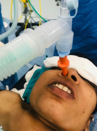
Inj.Glycopyrrolate 0.2mg intramuscularly was given 30 mins before induction in preoperative room, xylometazolin 0.1% nasal drops were given in both nostrils. Inj.Dexamethasone 0.2mg/kg IV and Inj.Hydrocort 2mg/kg IV given preoperatively. Difficult airway cart was prepared and toxic dose of local anesthetic was calculated. She was nebulised with topical lignocaine 4% 2ml followed by spraying of the base of tongue and posterior pharyngeal wall with 10% lignocaine before procedure. Inj. Dexmedetomidine 0.4mcg/kg IV given before procedure. Superior laryngeal nerve block were given with inj.Lignocaine 2% 1.5ml on both sides & transtracheal block given with inj.Lignocaine 2% 4ml. Before insetion of fiberoptic bronchoscope patency of the nostrils were checked. Patient was oxygenated with nasal airway with 6 mm connector from opposite nostril by bains circuit (Figure 3). Awake fiberoptic nasal intubation was done using spray as you go technique with maintaining cervical spine stability throughout and intubation was successful in first attempt. After securing the airway, intravenous induction was done with Inj.Propofol 2mg/kg IV and Inj.Vecuronium 0.1mg/kg IV and patient was taken on mechanical ventilator. Inj. Fentanyl 2mcg/kg IV & Inj. Paracetamol 15 mg/kg IV given as an analgesia. Surgery was lasted for 2 hours. Blood loss was minimal. Patient was hemodynamically stable throughout the procedure. She was reversed with Inj.Neostigmine 0.05mg/kg IV and Inj. Glycopyrrolate 8mcg/kg IV and extubated uneventfully. She was shifted to postoperative care unit. Postoperative analgesia was maintained with inj. Paracetamol 15 mg/kg IV TDS.
Discussion
Achondroplasia is the most common cause of dwarfism caused by gain of function mutation in fibroblast growth factor receptor 3 (FGFR3) gene. 80% mutations are the result of sporadic mutation while 20% are autosomal dominant. Incidence is approximately 0.5-1.5 in 10,000 newborns.1 Females are more affected than the male.2 Clinically, characteristic symptoms of disproportionate dwarfism is relative large head, midfacial hypoplasia. Achondroplasia affects multiple systems, primary or secondary to consequences of other organ systems affected.
A general recommendation regarding the ideal anaesthetic technique cannot be given, as both general and regional anaesthesia present potential problems.3 Therefore, individual decisions are necessary.
Problems during general anesthesia includes Excessive anxiety,4, 5 difficult intravenous access, difficult Laryngoscopy and endotracheal intubation6, 7 and difficult mask ventilation,8, 9 risk for cervico-medullary compression or rather spinal cord ischemia (reported sudden death events), ten-fold increased cardiovascular risk – with a maximum between 25 and 35 years of age,10, 11 eight-fold higher obesity rate potentiating the effects of the existing problems and increased incidence of sleep apnea (obstructive and/or central)9, 10 – rarely secondary pulmonary hypertension, restrictive lung diseases already at an early age,8, 9 chronic respiratory infections1 tendency toward hypersalivation. Increase incidence of gastro-oesophageal reflux disease in dwarf patients therefore there is a risk of aspiration.
Despite of all these problems during general anesthesia, we preferred general anesthesia over regional, due to the anatomical changes in the spine and the craniocervical junction as well as an increased incidence of hydrocephalus, spinal anesthesia is relatively contraindicated.
Neuraxial regional anesthesia is considered to be technically difficult due to narrow spinal canal/stenosis, reduced epidural space, kyphoscoliosis, vertebral body deformities.12 There are chances of potential neurological failures after regional anesthesia. In some cases, epidural anaesthesia was carried out successful. There are reports about accidental dural punctures, increased risk of venous puncture,13 difficulties in advancing the catheter, irregular or unpredictable spread of anesthesia.13 Epidural anesthesia should be preferred because of the possibility of titration. The conus medullaris is often positioned lower than usual.14 In most cases, injection of local anaesthetic into the caudal canal is easier in the case of paediatric patients.
Also spinal anesthesia even when applied successfully,15 carries the risk of failure of block as a result of unpredicted spread of the subarachnoid block due to anatomical deformity of spine.
While performing awake fiberoptic intubation, it is important to allay the anxiety of patient as these patients are highly anxious, we gave dexmedetomidine intravenously before procedure with vitals monitoring. Dexmedetomidine is a highly selective alpha 2 adrenoreceptor agonist and has a several unique properties, including sedation, anxiolysis, analgesia, hemodynamic stability and easy arousability. Dexmedetomidine demonstrates minimal respiratory depression16 even at a higher doses and also decrease salivary secretions, which are desirable during awake FOI.
Greatest care is required to prevent damage caused by positioning in case of anatomical abnormalities like khyphoscoliosis, lower limb valgus deformity, joint contractures and some patients are not able to lie supine.17 There are incidence of brachial plexus injuries during positioning in these patients. Case reports on damages caused by positioning do exist (e.g. two cases of brachial plexus palsy).18 Therefore we kept pillows underneath bilateral lower limb as a patient had joint contractures.
Compared to the size of the body, the head is relatively large & because of increase in body surface area they are more prone for hypothermia. Therefore, temperature monitoring and prevention of hypothermia is important in these patients.
Therefore in our case, we planned awake fiberoptic intubation as she had predicted difficult airway and to avoid neurological injury due to cervical spine extension during laryngoscopy, with all preoperative preparation for difficult mask ventilation and difficult intubation.


