Introduction
The breakage of an epidural catheter is a rare event which is encountered during both insertion and removal of catheter during epidural anesthesia.1 If the severed part is inert and if patient is asymptomatic with no complications, then its surgical removal is not recommended and it is left in the patient permanently.2
We encountered three cases of epidural catheter breakage over a period of one year.
Case 1
A 60yr old male, was scheduled for bilateral hernioplasty. After informed written consent, patient was planned for lumbar epidural with EPI CATH ® SFT-Romsons. The procedure was performed by a second year resident. Patient was positioned in left lateral. After local infiltration in the L2 L3 interspace, an 18 G Tuohy needle (Romsons) was used to locate the epidural space using the loss of resistance to air technique. The space was located at 6 cm depth and an 18 G epidural catheter (Romsons Epicath) was introduced. Blood was noticed in the catheter as it crossed the tip and catheter was gently withdrawn. It was observed that 1 cm of the distal end of the catheter was missing. So, procedure was abandoned and case was further proceeded with spinal anesthesia. Post operatively the patient was explained about the catheter breakage and reassured.
Case 2
A 75yr old male, ASA III scheduled for abdominal rectopexy. After informed written consent, patient was planned for lumbar epidural along with general anaesthesia. L2 L3 space was chosen and epidural space located with 18 G Tuohy needle and catheter was inserted. A third year resident performed the procedure. Resistance was felt when 18 G radio opaque catheter was introduced so it was decided to relocate, catheter was withdrawn with needle in situ and noticed that 2 cm of the distal end was missing in catheter. So, case was further proceeded with general anaesthesia.
Case 3
A 55 yr old male, ASAII scheduled for bilateral hernioplasty. After informed written consent, patient was planned for lumbar epidural. L3 – L4 epidural space was identified using 18 G Tuohy needle and catheter inserted.
As there was significant resistance during insertion the catheter was withdrawn and saline was planned to be injected to facilitate insertion, however the catheter tip was found to be sheared at the same location. Further case was proceeded with spinal anesthesia.
Each patient was explained about further management and complications but they were not willing for any investigations. So they were planned for scheduled follow up.
Discussion
Epidural catheters are made up of various materials including nylon, polyethylene, polyurethane and polyamide.3 Among these polyurethane catheters are less fragile even when traumatized.4 An ideal catheter should be flexible and disposable, radio opaque and have stretching capacity.
The breakage of epidural catheter may be due to various factors like kinked, knotted or curled catheter in epidural space, which causes it to stretch or shear, catheter trapping or compression between vertebral spinous processes,5, 6 impairment in catheter flexibility, manufacturing defects,7 injury of catheter by Tuohy needle, applying excessive force during removal.8
Olivar9 et al suggest that catheter be never withdrawn from the needle after it is passed through it, and to always withdraw needle and catheter as a unit, or the needle to be withdrawn first.
Even though it is common knowledge that catheters should be never withdrawn through the needle, the practice is still observed especially among residents. The possible reasons include reluctance to remove the needle and repeat the procedure especially in difficult cases. Performance anxiety and the possibility of repeating the procedure might appear as lack of the skill to peers and seniors deter them from removing the needle and reinserting. In high volume operating theatres like ours, residents do not get ample time to repeat procedures and usually taken over by the consultant at the sign of first difficulty. Prior experience significantly influences cognitive processes and skill based performance. Most of the residents were using PORTEX® Epidural Catheters-smiths medical till then and were used to gently withdrawing the catheter through the needle and use various techniques like slight advancement of the needle, injecting saline through the needle and then reinserting the needle to facilitate catheter introduction.10 Whereas the PORTEX® Epidural Catheters were flexible and the needle tip does not shear the catheter easily, the Romsons do. This highlights the importance of strictly adhering to the manufacturer’s recommendations while using medical devices.
Some more factors to consider avoiding breakages are as follows. Catheters may shear from imperfections such as nicks or barbs on the bevel of an unsharpened needle.11 Manipulation of catheter within the patient can cause catheter to become looped, trapped or knotted from curling back on itself when deflected by anatomical structures.11
Manufacturing defects can rarely cause catheter fractures.12 Catheters should be examined prior to insertion. Maximum length of the catheter within the epidural space should not exceed 5 cm. Epidural catheters should not be sutured to the skin which might cause breakage and a well trained person should perform catheter removal without using excessive force or tools like forceps.13 If difficulty is encountered during catheter removal, it’s suggested that the efforts at removal be stopped for 15-30 minutes.14
If the broken piece is symptomatic, it should be surgically removed as soon as possible. If the broken fragment is small, unable to detect, sterile and inert and if patient has no neurological complaints it can be safely left in place.7, 14
Considering these factors, in our case it may be thought that tuohy needle has cut the catheter. And since it’s a small undetectable fragment it can be safely left in place with periodic follow up of the patient.
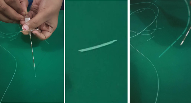
We tried to examine EPI CATH ® SFT- Romsons in vitro, (Figure 1) when we introduced the catheter we observed that when it crosses the needle tip the catheter tends to break or felt resistance at a specific area and gets shear off. The sharp tip of the Tuohy needle and the relatively rigid catheter as compared to PORTEX® Epidural Catheter might be the reason.15, 16
Patients were followed up for a period of one year and found to be asymptomatic without any complications.
Conclusion
Guidelines for insertion and removal of catheters should be followed to prevent shearing. Catheters should also be manufactured with materials that are high tensile strength, more resistant to breakages or shearing off. The presence of fragment should be documented and communicated to patients and surgeons, since neurological symptoms can develop months are years later. Therefore patient should be reviewed regularly and if symptoms develop, imaging studies and surgery are advocated.

