Introduction
Head fixation is a mandatory step in nearly all craniotomies for brain tumors' excision, neuronavigation surgeries and cerebral vascular interventions. Various techniques have been described for head fixation; starting from just taping the head firmly on a ring bellow or using a horse-shoe but these techniques were not satisfying for most of surgeons due to some preserved head mobility during craniotomies.1,2,3,4,5
Mayfield skull clamp (Figure1) was introduced by Frank Mayfield and George Kees in 1973 as a head holder during delicate intracranial procedures by application of sterile pins deep to the periosteum at two opposite sides of the head.6 But, insertion of these pins results in a sharp and intense noxious stimulus which results in a severe hemodynamic pressor response which in-turn may be hazardous for patients with disturbed autoregulatory mechanisms, increased intracranial pressure or cerebral va scular lesions.7,8
Many strategies have been reported to blunt this undesirable pressor effect: bolus injection of opioids, deepening level of anesthesia, infiltration of pins sites with local anesthetic drugs prior to its application and using of α2 - adrenoceptor agonists, but all of these strategies have been evidenced to have undesired side effects.9,10,11,12,13
Magnesium sulphate (MgSo4) which has been used widely by anesthetists, gynecologists and cardiologists as an anticonvulsant and antiarrhythmic has been reported to have analgesi c benefits through its N-methyl-D-aspartate (NMDA) receptor blocking effect beside its calcium channel blockade and reduction of catecholamine release.13,14,15
Many studies demonstrated the efficacy of MgSo4 in reducing either intraoperative anesthetic and opioid consumption or postoperative pain in different surgeries.13,16,17 But this prospective, double blind, and randomized study is the first to evaluate the effect of MgSo4 on attenuation of hemodynamic pressor activity after head clamp application during craniotomies.
Materials and Methods
This randomized, double blind, and prospective study was done in Neurosurgical department at ElS ahel Teaching Hospital in Cairo from February 2016 till august 2017. After approval of Hospital ethical Committee, written informed consent were signed by all enrolled patients before entry. All adult patients aged from 18 to 60 years of both genders, ASA physical status I and II scheduled for craniotomies were assessed to enter this study.
Patients with history of MgSo4 consumption or allergy, renal disease, hepatic or endocrine disorder, cardiovascular dysfunction, calcium channel blocker intake or drug abuse were excluded from this study.
Seventy one patients were assessed preoperatively, sixty of them were enrolled and assigned in two groups (n =30 each) using computer generated tables, random numbers were printed on previously prepared sealed envelopes to ensure blindness. For every patient, the envelop was opened by an independent technician to prepare 100 ml bottle of either regimens then gave it to the responsible anesthetist who was blind to its content.
Group M received 50 mg/kg MgSo4 in 100 ml 0.9 sodium Chloride 15 minutes prior to anesthesia induction over 15 minutes. Group S received 100ml 0.9% Sodium Chloride over the same period with the same rate.
Anesthetic technique
While in operating room, all patients were connected to all routine monitors: electrocardiography (ECG), pulse oximetry, non-invasive blood pressure monitor and peripheral nerve stimulator. All patients received midazolam 0.03 mg/kg intravenously as a premedication then the test solution was started over 15 minutes with a simultaneous infusion of 8 ml/kg normal saline as a preload. After insertion of an arterial cannula through radial artery, anesthesia was conducted using 2µg /kg fentanyl intravenously, thiopental 5mg/kg and followed by 0.6 mg/kg atracurium. After intubation with the proper sized armored endotracheal tube, anesthesia was maintained using isoflurane 1.1.5 minimum alveolar concentration (MAC) with oxygen: air (50:50) and an infusion of atracurium 0.01-0.15 mg/kg/minute was started. Ventilation was started according to guidelines to achieve end-tidal Co2 (ETCo2) between 35 to 40mmHg. All patients were managed according to our protocols till the end of surgery.
All the patients' monitoring outcomes were recorded continuously according to our hospital protocols, but our primary study outcomes were mean blood pressure (MAP), heart rate (HR) and the need for a bolus dose of fentanyl after pins insertion. MAP and HR were recorded just before head pins insertion (Pin pre) then at the time of pins insertion(Pin0) , after 60 seconds, 5 minutes, 10 minutes, 20 minutes and 30 minutes (Pin60'', Pin5', Pin10',Pin20' and Pin30' respectively).
Statistics
Sample calculation was done based on previous studies with a pow er of 80% and a type 1 error of 5%, at least 50 patients were needed to detect 20% difference between groups in blood pressure and heart rate. We increased number in each group to 30 patients to increase strength of the study. All data were collected on a separate sheet for every patient then gathered in an excel sheet using Microsoft excel (2010).
Statistical analysis was done using Statistical Package for Social Science (SPSS) IBM V.21. Both groups were compared for age, gender, weight, mean MAP and mean HR. student t -test was used for continuous variables as age and MAP but Chi-square test was applied to compare categorical variables as gender. P -values less than 0.05 were considered statistically significant.
Results
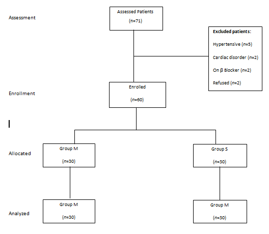
Seventy one patients were eligible for this study; sixty patients (84.5%) completed this study. Eleven patients were excluded because of hypertension (n =5), cardiac problem (n =2), receiving β- blocker (n =2) and patient's refusal (n =2). Data from excluded patients were discarded. (Figure 2)
Regarding demographic data; mean age of patients in group M was 50.07(±10.37) years while in group S was 51.13(± 8.6) years with no significant difference (p =0.669). Mean weight was 62.93(±14.12) kg in group M while in group S it was 63.17 (±12.92) kg, the difference was not significant (p =0.842). Fourteen male patients and sixteen females in group M were compared to sixteen females and fourteen males in group S with no significance between both groups (p =0.605). Time from induction till pins insertion in both groups showed no significance difference (20.83 ±3.6 vs 21.27± 4.79 minutes) with (p =0.691). (Table1)
Mean HR just before pins insertion (pinpre) was 74. ±5.91/min in group M and 75.47 ±5.21/min in group S without significance difference (p =0.312) while at insertion time (Pin0) it started to show significance difference (p ˂0.001) with a mean of 82.47± 4.49/min in group M and 104.24 ± 6.2/min in group S. In the subsequent reading at 5min, 10 min and 20min, mean HR showed the same significance (p ˂0.001). After 30 minutes (pin 30'), mean HR was 77.4± 3.83 /min in group M and 79.23 ±5.19/min. in group S with statistically insignificant value (p = 0.124). (Table 2)
Regarding mean MAP before pins insertion (pinpre) it was 77.27 ±6.55 mmHg in group m and 76.97 ±7.3 mmHg in group S which was statistically insignificant (p =0.861). While at time of insertion (Pin0) it was 80.5 ±6.45 mmHg in group M and 95.73 ±6.68 mmHg in group S with significant difference (p ˂0.001). At the following check points (at 60 seconds, 5min, 10 min and 20min), it showed the same significant difference (p ˂0.001). But 30 minutes after pins insertion, mean MAP was 74.53± 5.26 mmHg in group M and 76.73 ±4.91 mmHg in group S with no significant difference (p =0.1). (Table 3)
Another comparison was don e inside each group between pinpre, pin0 and pin60'' measures. At time of insertion, group M showed insignificant increase in mean MAP and mean HR (80.5 ± 6.45 mmHg and 76.7 ± 5.98 / min) with p-values of 0.06 and 0.08 respectively, while in group S both showed significant increase (p ˂0.001). After 60 seconds, both groups showed significant increase in both variables compared to pre-insertion measure with p ˂0.001 in each.
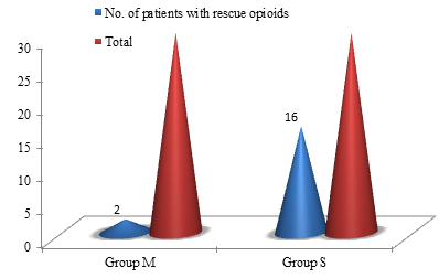
Two patients in group M (6.7%) versus 16 patients in group S (53.37%) received bolus dose of fentanyl due to sudden rise of HR and MAP 20% or more above pinpre measure (p ˂0.001). One patient in group S showed infrequent ventricular arrhythmia (1-3/min.) after pins insertion which did not necessitate any medical intervention. (Figure 3)
Table 1
Demographic data
Table 2
Comparison between groups M and S for Mean heart rate (HR)/ minute
Table 3
Comparison between groups M and S for mean arterial blood pressure (MAP) in mmHg
Discussion
Head fixation using Mayfield clamp has become an essential step during craniotomies worldwide. The manual insertion of its pins into the periosteum through scalp layers results in a sudden and severe noxious stimulus which leads to potential pressor response including sudden rise in MAP and HR which may – in turn- be detrimental for patients with coexisting cardiac disorder, increased intracranial pressure (ICP), intracranial vascular disease or disturbed auto-regulatory mechanism.
To blunt this response, many strategies have been described. One of these was the deepening of the level of anesthesia prior to pins insertion which may be unacceptable in many patients with cardiac comorbidity. Bolus injection of opioids as a rescue dose had been claimed for the increase of cerebrospinal fluid pressure which may be hazardous in space occupying lesions.4 Infiltration of the pins' sites with local anesthetic has been proved to blunt this pressor effect. With multiple trials done by some junior surgeons to apply pins in appropriate sites, defining the exact sites to block is sometimes uncertain. Intra- arterial injection into cerebral circulation has been recorded which may results in respiratory arrest or brain stem anesthesia.9
Magnesium sulphate (MgSo4) has been used widely anesthetists. Being a non-competitive N-Methyl-D-Aspartate (NMDA ) receptor antagonist and a calcium channel blocker, it has an analgesic effect by blocking nociceptive central sensitization. It has also an ability to reduce peripheral nociception by reduction of catecholamine release.18,19
This study demonstrated the efficacy of a single intravenous dose of MgSo4 (50 mg/kg in 100 ml 0.9 NaCl) prior to Mayfield clamp application during craniotomies in blunting the resultant hemodynamic pressor effect.
Many studies have been conducted to prove the efficacy of MgSo4 in reducing postoperative pain after different surgeries which showed a lot of controversy.13,16,17 Their opposed results was due to the multifactorial nature of pain perception postoperatively. Ethical, genetic psychological or even religious factors may cause these discrepancies.
Intraoperatively, MAP and HR are usually used as markers of pain in an anesthetized patient which necessitates a higher consumption of anesthetics and opioids. The efficacy of intraoperative effect of MgSO4 on intraoperative consumption of propofol and remifentanyl was studied by Ryu et al.20 who concluded that IV MgSo4 did not have any significant effect. This conclusion was opposed by Manaa and Alhabib21 who showed that MgSo4 decreased intraoperative consumption of fentanyl, propofol and rocuronium significantly, even with lower bolus and infusion doses they used. This can be explained by the different regimens in anesthesia they used, t his is why we used fentanyl in 2 µg / kg and 1-1.5 MAC of isoflurane in our patients. Our results showed the same significant reduction in fentanyl consumption after MgSo4 administration.
In present study, sudden increase in MAP and HR has been noticed at time of pins ' insertion with a mean rise of 3.2 mmHg and 76.7/min respectively in magnesium group versus 18.8 mmHg and 97.2/min in saline group with a significant difference between both groups. The peak rise was noticed after 60 seconds of pins insertion (8.7, 26.3 mmHg and 82.47, 104.27/min in both groups). This was the same peak time recorded by Arshad A et al.22 for this noxious stimulus to cause its maximal pressor effect. At the consequent check points, the same significant differences were found between both groups regardless the gradual decrease in different means.
However; at 30 minutes both groups showed no significance difference which can be explained by the normalization of hemodynamic response which fades away by time after sudden painful stimulus due to the natural neuromodulation mechanisms including depletion of neurotransmittors. Another explanation for this regression is the more depth of anesthesia which has been achieved at that time.
Despite of its calcium channel blocking activity, no severe hypotension has been noticed with magnesium administration before pins insertion, this was explained by the usage of 50 mg/kg which is lower than the minimum dose of MgSo4 proved to cause hypotension which was 60 mg/kg.19,23
Magnesium toxicity was claimed to cause cardiac arrest with serum concentrations above 2.5-5.0 mmol/ liter, but according to Goral N et al24 administration of bolus dose of 50mg/kg followed by an infusion of 20mg/kg/hr didn't reach this toxic serum level. During this study we administered lower total dose of MgSo4, so we didn't monitor Mg serum level.
Apart from being double-blinded and randomized study with an appropriate sample size, two limitations have been realized; first, comparison of MgSo4 to one or more of the evidence based strategies will be beneficial. Second, monitoring of intracranial pressure as an outcome could be helpful but we faced some logistic difficulties during this work.
In conclusion, our study demonstrated the efficacy of a single intravenous dose of MgSo4 (50 mg/kg in 100 ml 0.9 NaCl) in attenuating the hemodynamic pressor effect resulting from the application of Mayfield clamp during craniotomies without any adverse effects.


