- Visibility 85 Views
- Downloads 7 Downloads
- DOI 10.18231/j.ijca.2021.103
-
CrossMark

Integrated oxygen therapy consisting of non invasive ventilation and nasal cannulae in respiratory failure secondary to COVID-19 pneumonia: Case series
Introduction
COVID-19 pneumonia caused by SARS-CoV-2 is a rapidly evolving global emergency that has swept off significant numbers of human lives and continues to do so in the presence of very limited evidence based recommendations for its management. Patients with moderate to severe COVID-19 develop hypoxemic respiratory failure demonstrating an uncoupling of degree of hypoxia and respiratory distress. Profound degrees of hypoxia have been observed with little or no distress putting the conventional management consisting of an early intubation in question.[1], [2] In this case series, we attempted an integrated oxygen strategy utilizing non invasive ventilation with standard oxygen therapy (SO) via nasal cannula. By increasing oxygen concentration in the NIV masks, oxygenation improved as well as the respiratory rate decreased, hence decreasing work of breathing. Both the patients were managed with successful outcomes, without the need of intubation. A similar dual oxygen therapy has been implemented by Kumar et al in non-covid critically ill patients with successful outcomes.[3] Implementation of this strategy helped us in avoiding intubation and preserved much needed critical care ventilators during times when our nation was hit hard by the second wave of pandemic.
Case 1
A 52 yr old male got admitted in ICU with complaints of fever and cough for 13 days. He was known hypertensive for the past 5 years, on treatment. BMI was around 35 kg/sq.cm. He tested RTPCR positive for Covid 19 on day 5 of illness. On presentation, he was maintaining oxygen saturation of 85% on room air, associated with a respiratory rate of 32 per minute. His chest X.
ray was suggestive of bilateral pulmonary infiltrates. Initially, he was administered oxygen therapy via non rebreathing mask @10 litres/minute, which had to be increased upto 15 litres/min. On day 6 due to failure to maintain adequate saturation (88-90%), he had to be administered non-invasive mechanical ventilation via CPAP 10 cm H20 and Fi02 0.6.
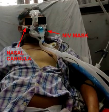
Eventually, he needed to be put on Bi-level positive pressure ventilation (BiPAP) with pressures 14/6 mm Hg (iPAP/ePAP) and Fi02 of 0.7, to which he responded partially by maintaining Sp02 90-92%. Around day 9, the patient developed respiratory failure on BiPAP, with increased spontaneous breathing rate and falling oxygenation. At this point of time, the patient was administered oxygen therapy with nasal cannula @ 10 litres/min along with BiPAP with PSV 16 cm H20 and PEEp 8 cm H20 @ 0.9. ([Figure 1]) After around an hour, the patient's respiratory rate reduced, Sp02 improved to 98-99% and the patient became comfortable. Patient was administered on the same pressure support for the next 3 days, after which we could taper the flow of the nasal cannula to 4l/min and put him off the nasal cannula. The ventilatory support could be tapered to NRBM over the next few days, with the patient maintaining saturation on NRBM @10 litres/min. Eventually, the patient was discharged on room air on day 27 of admission. [Table 1] depicts serial Arterial blood gas analysis of the patient during ICU stay. Patient’s medical management included antibiotics meropenem, steroids and prophylactic dose of clexane.
|
|
Day 0 |
Day 4 |
Day 12 |
Day 13 |
Day 17 |
Day 20 |
Day 23 |
Day 27 |
|
|
NRB M@1 5l/mi n |
CPAP 10 @ 0.8 |
BiPAP 16/8 @1.0 |
BiPAP 16/8 @ 0.9 With NC @6l/min |
BiPAP 16/8 @ 0.8 With NC @6l/min |
BiPAP 14/6 @0.7 |
NRBM @ 10 litres/mi n |
Venturi mask@0. 35 |
|
Sp02 |
92-93 |
92-94 |
89-90 |
94-95 |
96-98 |
96-97 |
95-97 |
94-96 |
|
pH |
7.42 |
7.42 |
7.46 |
7.41 |
7.39 |
7.45 |
7.39 |
7.38 |
|
Pa02 |
84.1 |
74 |
55 |
79 |
92 |
90 |
94 |
89 |
|
PaCo2 |
32 |
36 |
29 |
36 |
39 |
38 |
32 |
36 |
Case 2
A 45-year-old male with history of type 2 DM, hypertension and sarcoidosis on medical management, presented with high grade fever, cough and shortness of breath for 6 days. He tested positive on RT PCR for SARS-CoV 2 and was shifted to COVID ICU. On admission, he was febrile, with a pulse rate 121/min, blood pressure 126/89 mmHg and oxygen saturation 88% on room air. The oxygen saturation was optimized to 92% on non rebreather mask @15 L/min. All blood investigations were essentially within normal limits, while CRP was 10 mg/dl and D dimer was 4.5 mcg/ml. Arterial blood gas analysis on admission, on NRBM @15l/min revealed Pa02 84.2, PaCo2 41.0 and pH 7.39. Chest x-ray revealed bilateral pulmonary infiltrated. HRCT chest revealed ground glass opacities, with CT severity score of 18/25 suggestive of severe disease. Over the next few days, his oxygen requirement kept on increasing, with upscaling of ventilatory support to CPAP @10 cm H20 and Fio2@0.6.At day 12 of admission, due to the worsening hypoxemia and persisting tachycardia and tachypnea, he was administered BiPAP, with 16 /8 cmh20 (iPAP/ePAP), with Fio@0.9.We were unable to maintain Spo2 above 84-85%, and other clinical variables like respiratory rate and pattern continued to deteriorate. Subsequently, we administered oxygen therapy @10l/min via nasal cannula as well as BiPAP. Sp02 maintained above 92 % for the next few hours. After around 12 hours, clinical improvement was observed in terms of reduction of respiratory rate, diminution of accessory respiratory muscle use and sp02 maintaining above 96 percent. . The patient was kept on the same settings for the next 96 hours, following which the oxygen requirement was reduced.
Eventually, gradual weaning was successful, and the patient was discharged @ day 29 of illness as he was maintaining saturation 96-98% on room air. [Table 2] depicts serial ABG values of the patient during the stay. Patient received injection remdesivir. Injection piperacillin tazobactam, inj methylprednisolone, inj clexane during his ICU stay.
|
|
Day 0 |
Day 1 |
Day 9 |
Day 9 |
Day 10 |
Day 14 |
Day 23 |
Day 25 |
|
|
NRB M@15 l/min |
CPAP @10 Fi02 0.6 |
BiPAP 16/8 @0.9 |
BiPAP 16/8 @ 0.9 With NC@ 10l/min |
BiPAP 16/8 @ 0.9 With NC@ 10l/min |
BiPAP 14/6 @0.7 |
NRBM @ 10l/min |
Ventu ri mask @0.3 5 |
|
Sp02 |
94-95 |
95-96 |
89-90 |
94-95 |
96-98 |
96-97 |
95-97 |
94-96 |
|
pH |
7.41 |
7.39 |
7.46 |
7.41 |
7.42 |
7.45 |
7.39 |
7.38 |
|
Pa02 |
72.1 |
74 |
55 |
73 |
98 |
90 |
94 |
89 |
|
PaC02 |
38 |
41 |
32 |
36 |
41 |
34 |
37 |
35 |
Discussion
The ventilatory management of acute respiratory syndrome secondary to COVID-19 pneumonia remains a controversial subject. Gattinoni et al. described two different phenotypes in COVID-19, with only 20-30 per cent demonstrating a typical ARDS like pattern, suggesting that they might need to be managed differently.[4] Recent review of literature also revealed that the timing of intubation has a negligible impact on the mortality and morbidity of critically ill COVID patients and perhaps a wait and see approach may be justified.[1], [2] In this case series, we attempted an integrated oxygen therapy consisting of NIV along with oxygen therapy via nasal cannulae in patients who failed to maintain oxygenation on BiPAP alone.
One of the primary causes of failure to maintain oxygenation during NIV is the interface for NIV and Ventilator surface which comprises apparatus dead space and accentuates carbon dioxide rebreathing.[5], [6]
Therefore, by increasing the oxygen flow rate this therapy helped in reducing CO2 rebreathing.
High minute ventilation in patients of hypoxemic respiratory failure on NIV has been associated with higher mortality.[7] This therapy might have proven helpful by suppression of the hypoxic respiratory drive as was evident by subsidence of hyperventilation in both patients.
We propose the following reasons behind improved oxygenation following this integrated oxygen therapy:
Increased oxygen concentration inside the mask
Prevention of carbon dioxide rebreathing
Washout of anatomical dead space in the nasopharynx
Suppression of the hypoxic drive
Delivering oxygen therapy via nasal cannula helped us minimize the pressure support in the context of NIV. Therefore, this therapy might help in preventing barotrauma and pneumothorax in the susceptible patients of Covid-19 pneumonia.
Careful patient selection is a prerequisite before attempting this approach since patients with high inspiratory pressures or showing high inspiratory drive should be precluded and considered for intubation. Another prerequisite is vigilant monitoring as well as ready availability of the intensivist since rapid decompensation or increase in the work of breathing would necessitate intubation. Development of spontaneous pneumothorax and pneumomediastinum is another complication one needs to be watchful about, which has been observed with higher incidence in this patient population.[8]
This proved to be a saviour technique for us when we were amidst a prevalent oxygen crisis due to the second wave of pandemic, and could not administer high flow nasal cannula therapy to our patients. By avoiding intubation, we could preserve much needed critical care ventilators during the time of crisis and use them in patients in whom intubation was the only resort.
Conclusion
Through this case series, we intend to propose an integrated oxygen therapy consisting of non-invasive ventilation and SO by nasal cannula for ventilatory management of patients in respiratory failure secondary to COVID-19 pneumonia and hence avoid intubation. This might help in preservation of critical care ventilators and help improve patient outcomes in the areas hit hard by the pandemic. We suggest multicenter studies with larger sample sizes to be undertaken as soon as possible to validate our findings.
Source of Funding
None.
References
- AC Hernandez-Romieu, MW Adelman, MA Hockstein, CJ Robichaux, JA Edwards, JC Fazio. Timing of intubation and mortality among critically ill coronavirus disease 2019 patients: a single-center cohort study. Crit Care Med 2020. [Google Scholar] [Crossref]
- P Rola, J Farkas, R Spiegel, C Kyle-Sidell, S Weingart, L Duggan. Rethinking the early intubation paradigm of COVID-19: time to change gears?. Clin Exp Emerg Med 2020. [Google Scholar] [Crossref]
- A Kumar, L Kumar, C Sinha, N Kumar, U K Bhadani. Dual oxygen therapy in patient on bilevel positive airway pressure prevented invasive mechanical ventilation. Indian J Crit Care Med 2017. [Google Scholar]
- L Gattinoni, D Chiumello, P Caironi, M Busana, F Romitti, L Brazzi. COVID-19 pneumonia: different respiratory treatments for different phenotypes?. Intensive Care Med 2020. [Google Scholar] [Crossref]
- GP Schettino, S Chatmongkolchart, DR Hess, R Kacmarek. Position of exalation port and mask design affect CO2 rebreathing during non-invasive positive pressure ventilation. Crit Care Med 2003. [Google Scholar]
- J Gonzalez, T Sharshar, N Hart, K Chadda, J C Raphae L, F Lofaso. Air leaks during mechanical ventilation as a cause of persistent hypercapnia in neuromuscular disorders. Intensive Care Med 2003. [Google Scholar]
- L Gattinoni, S Coppola, M Cressoni, M Busana, S Rossi, D Chiumello. COVID-19 does not lead to a “typical” acute respiratory distress syndrome. Am J Respir Crit Care Med 2020. [Google Scholar]
- TS Elhakim, HS Abdul, CP Romero, Y Rodriguez-Fuentes. Spontaneous pneumomediastinum, pneumothorax and subcutaneous emphysema in COVID-19 pneumonia: a rare case and literature review. BMJ . [Google Scholar] [Crossref]